Anatomical Descriptions of the Snow Leopard
Project Title: Anatomical descriptions of the snow leopard
Start Date: 2018
Lead Researcher(s): Dr. Beth Townsend, Dr. Heather Smith and Dr. Scott Echols
Contributing Researchers: Dr. Julie Barnes, Dr. Erika Crook, Dr. Nancy Carpenter, Shaghayeagh Assar, Madelyn Durhman, B. Adrian, S. Marsh, S. Levy, R. Hassur, S. Nagy, H. Mohamed, Dr. A. Grossman
Project Purpose: To define and publish the anatomy of the snow leopard (Enteroctopus dofleini). The goal is to create numerous publications highlighting special anatomic features of the snow leopard–especially those that explain special adaptations. Additionally, we hope to publish the whole animal anatomy.
Funding Needed: Completed.
Special Notes: Two full grown snow leopards (one male and one female) have been entered into the study.
Publications Produced: Yes
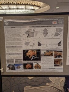
Townsend KEB, Smith HF, Manfredi KR, Assar S, Durhman M, Echols MS. Head and neck musculature of the snow leopard (Panthera uncia). American Association of Anatomy Annual Conference, Washington DC, 2023.
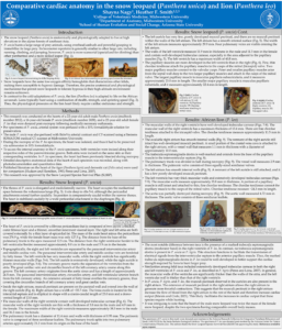
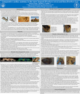
Nagy S, Smith HF. 2022. Comparative cardiac anatomy in the snow leopard (Panthera uncia) and lion (Panthera leo). Poster presentation at 2022 Society for integrative and Comparative Biology conference, Phoenix, AZ.
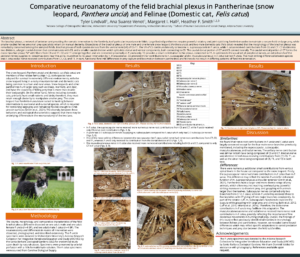
Lindvall T, Suarez-Venot A, Hall MI, Smith HF. 2022. Comparative neuroanatomy of the felid brachial plexus in Pantherinae (snow leopard, Panthera uncia) and Felinae (Domestic cat, Felis catus). Poster presentation at 2022 Society for integrative and Comparative Biology conference, Phoenix, AZ.
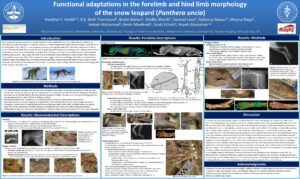
Smith et al. Functional adaptations in the forelimb and hind limb morphology of the snow leopard (Panthera uncia), Society for Integrative and Comparative Biology Jan, 2021.
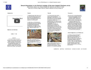
Marsh et al. 2020. (Snow leopard forelimb sexual dimorphism), 2020 National Veterinary Scholars Symposium: https://nvss2020-aavmc.ipostersessions.com/Default.aspx?s=8D-BE-29-BC-83-85-94-33-68-39-B3-FA-9D-32-AA-A8
Levy et al. 2020. (Snow leopard forelimb adaptations), 2020 National Veterinary Scholars Symposium: https://nvss2020-aavmc.ipostersessions.com/Default.aspx?s=D4-69-25-11-2D-0D-5F-E9-DA-0D-45-5B-A0-C5-B8-CE
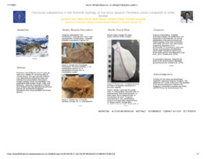
Smith HF, Townsend KEB, Adrian B, Levy S, Marsh S, Hassur R, Manfredi K, Echols MS. Functional Adaptations in the Forelimb of the Snow Leopard (Panthera uncia). Integrative and Comparative Biology. 1-15. doi:10.1093/icb/icab018.
Smith et al. 2021. ICB Snow leopard forelimb
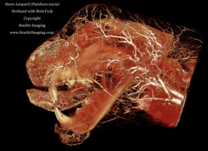
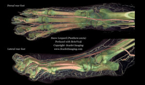
Hall MI, Lindvall T, Suarez-Venot A, Valdez D, Smith HF (2023) Comparative anatomy of the felid brachial plexus reflects differing hunting strategies between Pantherinae (snow leopard, Panthera uncia) and Felinae (domestic cat, Felis catus). PLoS ONE 18(8): e0289660. https://doi. org/10.1371/journal.pone.0289660
Australian Marsupial Anatomic Study
Project Title: Australian Marsupial Anatomic Study
Start Date: 2020
Lead Researcher(s): Dr. Justin Adams, Dr. Scott Echols
Contributing Researchers: To be determined.
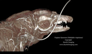
Project Purpose: To define and publish the anatomy of opportunistically available Australian marsupials. We will likely begin with brush tailed possums (Trichosurus vulpecula) and koalas (Phascolarctos cinereus). Part of the purpose is to determine how best to preserve, contrast perfuse and study marsupials many of which are in decline in the wild.
Funding Needed: To be determined.
Special Notes: The information gained from this preliminary study will be used to work towards endangered marsupials such as the Tasmanian devil (Sarcophilus harrisii) and even extinct species currently preserved in special collections.
Publications Produced: Not at this time.
California Sea Lion Anatomy Study
Project Title: California Sea Lion Anatomy Study
Start Date: 2020
Lead Researcher(s): Dr. Beth Townsend, Sea World, Dr. Scott Echols
Contributing Researchers: Justin A. Georgi, Ph.D., Ari Grossman, Ph.D., Margaret I. Hall, Ph.D., Christopher P. Heesy, Ph.D., Andrew Lee, Ph.D., Erin Simons, Ph.D., Heather Smith, Ph.D., Juan Rodriquez-Sosa, Brent Adria, M.F.A., Nora Wells
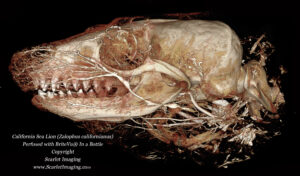
Project Purpose: To define and publish the anatomy of California sea lion (Zalophus californianus) heads.
Funding Needed: Fully funded.
Special Notes: This is designed to be a primary and pilot study. The primary study involves documenting the anatomy of the sea lion heads. Also, this study serves as a pilot for studying the anatomy of other large sea mammals such as whales and dolphins. Additionally, we hope to document the full anatomy of a sea lion if one becomes opportunistically available and there is dissection/documentation time available.
Publications Produced: Not at this time.
Sloth Anatomy Study
Project Title: Sloth Anatomy Study
Start Date: 2020
Lead Researcher(s): Dr. Ray Wilhite, Dr. Scott Echols
Contributing Researchers: To be determined.
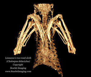
Project Purpose: To define and publish the anatomy of opportunistically available sloths. Since our first submission is a two toed sloth (Choloepus sp), this will be the first animal to be examined.
Funding Needed: Currently, fully funded for the first sloth only.
Special Notes: The information gained from this preliminary study will be used to work towards a compilation of information on sloth anatomy. Special thanks to Dr. Attila Molnar for his contribution.
Publications Produced: Not at this time.
Ferret and Mink Anatomic and Clinical Study
Project Title: Ferret and Mink Anatomic and Clinical Study
Start Date: 2021
Lead Researcher(s): Drs. Scott Echols, Nico Schoemaker, Heather Smith and Yvonne Van Zeeland
Contributing Researchers: Drs. Ari Grossman, Leigha Lynch and Carrie Veilleux
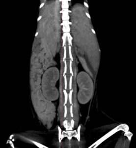
Project Purposes: 1. To define and publish the anatomy of ferret (Mustela putorius furo) and American mink (Neovison vison). 2. Develop clinically useful means to image and define key structures using advanced clinical CT in effort to better diagnose a variety of diseases common to ferrets. 3. Establish genetic markers specific to both species.
Funding Needed: Not determined yet.
Special Notes: This project is designed to result in numerous studies. The primary study involves documenting the anatomy of the common (pet) ferret and American mink. Both are commonly kept animals (ferrets as companion animals and mink in industry) and detailed anatomic accounts are lacking. We also will be concurrently evaluating different CT imaging (with and without contrast) protocols with the goal to better identify diseases that commonly plague ferrets. If possible, we will do the same with mink. Last, genetic sampling and analysis will be conducted on all animals.
Publications Produced: Not at this time.

